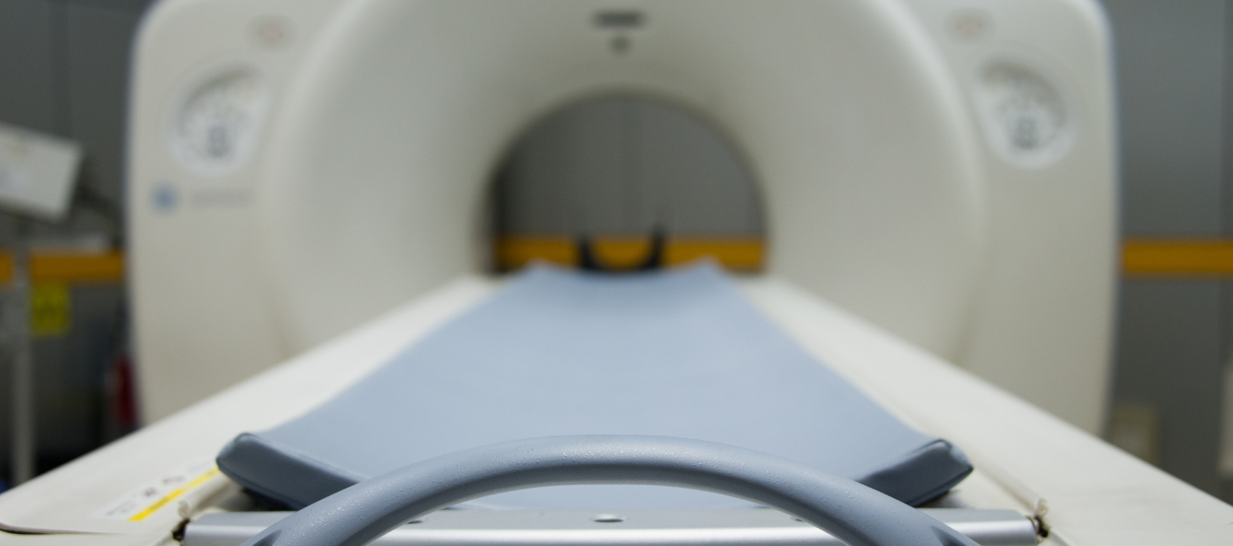
How to Prevent Patient Burns During MR Imaging

In this article, we review eight ways you can use to prevent patient burns during MR imaging. Watch the full explanation below in our YouTube video provided by Eric from Olympic Health Physics.

Eight Tips For Preventing Burns on MRI Patients

Download your free copy of the FDA’s poster on MRI Burn Prevention: Tips For Keeping Patients Safe to follow along with us. 

1. Screen Your Patients

The first process to implement that can prevent burns during MR imaging is to screen your patients before entering Zone 4. This includes screening for anything metallic, such as implants or medical devices. It’s a good rule to assume that anything unknown in or on your patient is not MRI safe. 

2. Screen Any Objects Going Into Zone 4

In addition to screening your patients, you also want to screen anything going into the MRI scan room. All objects entering Zone 4 need to be MR Safe or MR Conditional. If any MR Conditional objects enter the scan room, match them to the MR Conditional devices with your scanner. Remember that all metals, even non-ferromagnetic metals, have the potential to cause burns during MR imaging. 

3. Have Patients Change Into Hospital or Medical Gowns

Whenever possible, have your patient change out of their street clothes into a medical or hospital gown before entering Zone 4. This can prevent any metallic items, such a metallic fabrics, buttons, zippers, or embellishments, from unknowingly being exposed to MRI equipment. In addition, it prevents a patient from accidentally having metallic objects in their pockets and bringing them into Zone 4.

4. Ensure The Patient Isn’t Creating Conductive Loops

Next, you want to ensure that your patients aren’t creating any conductive loops themselves. For example, when a patient needs to be scanned with their arms over their head, you want ensure they don’t have their hands clasped. Your patient also shouldn’t cross their arms or feet. This avoids creating magnetic loops, which helps with burn prevention.

5. Use The Manufacturer Provided Padding

You want to use the manufacturer provided padding to pad the sides of the bore or in between the patient to insulate the patient. While you can use sheets and pillows, they should only be used for patient comfort. All padding and insulation should use manufacturer-supplied padding. 

6. Cables Should Run In A Straight Line From The Scanner

Another way to prevent burning the patient is to run any cables to and from scanner in a straight line. Check that the cables running from the coil into the magnet are not forming loops.

7. Use The Lowest SAR In Normal Operating Mode

While operating in normal mode try to keep the lowest SAR possible. If you have an SA monitor, keep an eye on the SA level to ensure that you’re within limits. 

8. Stay In Communication With Your Patient

Remain in communication with your patient at all times. Stay in visual contact when possible and using an intercom for verbal communication is essential.

Ensure the patient has the squeeze ball and give them directions on how and when it’s appropriate to use before you start the scan. 

Some MRI suites don’t allow you to keep eyes on the patient the entire time based on the orientation of the control room. If that’s the case, have some other way that you can monitor the patient.

Stop the scan and investigate the possible cause if the patient does communicate with you and tell you that they feel burning or feel something heating up.

And that wraps up the eight different ways that we can try to prevent MRI burns in MRI departments. 

You can find the FDA’s MRI burn prevention poster by clicking here 

Don’t forget that we also provide a variety of courses, including MRI Safety for MRI Professionals.

If you have questions about MRI or MRI safety, feel free to drop us a note and we’ll be happy to take a look at your situation and see if there’s something that we can do to help you.

Click the image above to download the file

We perform physics testing for all makes and models of MRI scanners.  For a complete description of our physics testing, check out our MRI Physics Tests.  In addition to our MRI Physics Testing, we also provide MRI Safety Audits for facilities wanting a comprehensive MRI safety assessment.

Contact Us for a No Obligation Quote

253-254-6988

Patient Radiation Safety in CT: What You Need To Know

Patient Radiation Safety in CT - What You Need to Know

In this week’s video, Eric from Olympic Health Physics explains the 10 pearls of radiation protection of patients in CT, as recommended by the IAEA (International Atomic Energy Agency) for the Radiation Protection of Patients (RPOP).  This help will give some guidance on the best ways that we can employ to protect our patients when using computed tomography imaging. 

1. Perform The Scan Only If It's Indicated

Try to avoid any unnecessary exams.

Remember that the lowest radiation exposure to a patient is the radiation exposure that doesn’t occur. This is why it’s important to know that if there’s an exam that shouldn’t be performed, then we shouldn’t be scanning the patient.

2. Consider The Use of Alternative Imaging Options

You can use alternative modalities to potentially answer the same clinical question.

Ultrasound or MRI don’t use radiation and they could be alternatives depending on the diagnostic question that’s trying to be answered. If a patient can be imaged using ultrasound or MSI from a radiation dose perspective, then it can be an excellent option when those modalities are appropriate.

3. Always Check If Your Patient May Be Pregnant​

We don’t want to unknowingly image or do a CT on someone who is either pregnant or potentially pregnant. That’s the reason why you always need to verify the patient, whether or not they could be pregnant before doing their exam.

You can do this by having postings in the department that say, “Please notify the technologist if there’s a possibility that you’re pregnant”. Another option is to directly ask the patient if there’s any possibility that they could be pregnant.

4. Start Using Images With Some Noise Without The Loss of Diagnostic Information

Always check if your patient may be pregnant.

It’s important to know that high-quality, detailed, and crisp images look really nice, but they may not be necessary for answering the diagnostic question.

You could potentially use a lower radiation dose and still answer the diagnostic question. The trade-off is that we’re going to introduce some noise and potentially lose some image quality. However, that loss and image quality are not going to necessarily change the diagnosis or the outcome for the patient.

We want to make sure that you’re using the right radiation dose for answering the clinical question.

5. Use Indication-Specific CT Protocols For Each Body Part

We want to use indication-specific protocols where they’re applicable. For example, not every chest CT should be a full chest image. We could do a low-dose screening, a lung screening chest protocol, and also potentially do a low-dose nodule follow-up protocol.

We want to use very specific protocols for the type of exam that we’re trying to do. The reason is that oftentimes those very specific protocols can be acquired at a lower radiation dose to the patient.

6. Multiple Pass or Multiphase CT Should Not Be Performed Routinely

It’s important to know that multiple pass or multiphase CT should not be performed routinely. We want to try to limit the amount of passes that we make through the patient. Only do the passes that are actually necessary and potentially combine different protocols.

This is why multiphase CT studies can often be 2 to 3 times the amount of radiation dose as just a regular CT.

7. Adjust Exposure Parameters According To The Patient And Body Part

We want to adjust the technique that we’re using to image our patient for the actual patient size. First, we want to take into account the size of the patient and the exam. Then, adjust any technique that we’re using so  it’s specific to the patient and the body part that we’re imaging.

The reason for this is to get away from a one size fits all technique and move to very patient-specific techniques. That way, smaller patients receive lower radiation doses than larger patients.

8. Know Your Equipment, Including the AEC System

Another important aspect is to know your equipment. For example, understand how the AEC (Automatic Exposure Control) works or the tube current modulations and how that works for your system. 

How Does CARE Dose 4D Work?

If you’re scanning on a Siemens scanner, make sure you understand CARE Dose4D. And if you’re scanning on a GE scanner, make sure you know how AutomA works. This way you can use the scanner to the best of its ability and it will help you give the right radiation dose to your patient.

To learn more about the Siemens CARE Dose4D system, check out our previous post here. And for information on the GE AutomA system, you can find our latest post here

9. Use Good Technique

Use good technique when we’re talking about good technique. These are things like:

•  Making sure that your patient is iso-centered within the gantry. This is in both the lateral direction as well as the AP direction.

•  Confirm that the patient is iso-centered, particularly while using tube current modulation.

• Check that your scan length is covering only the anatomy that’s absolutely necessary. We don’t want to scan the diagnostic portion of the CT.

The one caveat to this is with a scout. When using a scout, it’s better to use a little bit longer of a scout than what you intend to cover with your diagnostic scan. Keep in mind that he diagnostic scan should be shorter than your scout.

10. Pay Attention To Radiation Dose Values

Don’t forget to pay attention to your radiation dose values, such as the pre-scan CT because you need to make sure that you’re falling within your pre-determined dose values and dose limits.

Click the image above to download the file

Review the doses that you’re using on a monthly basis to see if there are any adjustments that you can make. Consider things like your protocols, your techniques, and how you’re scanning patients. From this information, you can have a pretty good idea of what your doses should actually be, what they have been and what they should be for the equipment that you’re using as well as for what your radiologists are accustomed to looking at.

Now you know that there are ten different ways to protect your patients, reduce their radiation dose and make sure that they’re getting the right radiation dose for the type of exam that’s been ordered.

Click here to download your copy of the IAEA’s 10 Pearls of Radiation Protection of Patients in CT.

Our team is dedicated to ensuring that your facilities’ radiation safety program functions in accordance to regulatory standards, sound radiation safety principles, and most importantly serves to protect staff, patients, and the general public from the hazards associated with ionizing radiation. To this end, Olympic Health Physics can provide a physicist to your organization to fulfill the Duties of the Radiation Safety Officer. To learn more, check out our RSO Services or click the link below. 

Contact Us for a No Obligation Quote

253-254-6988

How To Use a Geiger Counter

How To Use a Geiger Counter

In this week’s video, Eric from Olympic Health Physics explains how to use a Geiger Counter. Throughout the video, he demonstrates the Ludlum 14C with a pancake detector. He provides an overview of how to test the battery, how to ensure the counter is working properly, and how to read the panel.

What Is A Geiger Counter?

A Geiger Counter or Geiger Meter is an electronic instrument used to detect and measure the amount of ionizing radiation levels. They are widely used in various applications, including radiation protection and safety, radiation dosimetry, and the nuclear industry. If you work in a Nuclear Medicine department, you likely have a Geiger Counter to measure radiation in different aspects, such as wipe tests, area surveys, or exposure rate measurements.

Three Essential Checks Before Using Your Geiger Counter

When you’re using a Geiger Counter, there are three things that you need to check before you start using it. Throughout this post, we will be showing the Ludlum 14C with a pancake detector. Please note that there are many different Geiger Counter makes and models, so check your user manual for specific criteria for your equipment. 

1. Is The Geiger Counter's Calibration Valid?

The first thing is you need to make sure that the Geiger Counter is in calibration. You need to look at the calibration label on it to make sure that it is still within calibration.

The calibration certificate usually includes the last date of calibration and the expiration date. The sticker also includes other useful information such as the model numbers, serial numbers, and efficiencies. 

Geiger Counter Calibration Certificate

2. Is The Geiger Counter Responding Appropriately?

The second thing that you need to do is make sure that the instrument responds to a radiation source.

On this specific instrument, we have a check source with a window that we can we can open. We can then put our pancake detector over the cesium button source located on the side of the instrument to ensure the instrument is actually responding to radiation.

3. Are the Batteries Operating Within the Test Range?

The last thing you want to do is check the batteries to ensure the batteries are operating within the battery test range.

To test the batteries, first remove the pancake detector from the front of the Geiger counter. Then, turn the Geiger counter on and turn the knob to the lowest range setting. Test the battery by depressing the battery button and looking to make sure that the indicator stays in the battery test range. As long as the indicator stays within the battery test range, the battery is operating effectively. 

Geiger Counter Battery Test Button

Understanding the Instrument Scale Readout

Understanding how to properly read the instrument scale readout is important to ensuring you have the correct information.

You’ll note on the Ludlum 14C Geiger counter instrument there are three different scales on the face of the survey meter. The first scale or the bottom scale is in milliroentgen per hour. You can also notice that the very bottom scale indicates “X100 ONLY”. The second scale or the mid-range scale is also in milliroentgen per hour. The upper scale that goes across the top  of the readout is in counts per minute.

Instrument Scale Readout

Below the instrument scale readout on the top of the meter are the various potentiometer values you can set for the Geiger counter. On this particular model, the Ludlum 14C, there are five potentiometer value setting options you can select. The value selected determines the multiplication factor for any reading that you see on the face of the survey meter. 

The bottom three options include X0.1, X1, and X10 coincide with the middle and top readout. X100 is used for the bottom readout only. And the X1000 option is used for the internal probe only. 

Potentiometer Value Options

To understand how the potentiometer options and scale readout work together, consider the following example:

You select the value of X1 from the options available. When you scan the radiation source with the pancake detector, the needle on the readout stops at blue line on the example readout photo. In this example, the appropriate reading would be on either the middle or top scale range, depending on what information you require. In addition, the potentiometer value is X1, meaning the multiplication factor is only one. Therefore, the value seen is the actual measurement. So for this example, the reading is either 4,000 counts per minute or 1.2 milliroentgen per hour.

Suppose you had selected X10 and the needle result was the same. You would them multiple your readout results by 10 to determine your final measurement reading. 

Sometimes on your survey meter, you may notice no deflection of the needle at all. If this happens, it can often be caused by having it set on too high of a scale. To fix this, flip down to the lowest potentiometer scale and you should start to see needle deflection. 

Audio and Speed

The Ludlum 14C Geiger Counter also offers two additional options: audio and speed.

To turn the audio on, flip the switch up and you will hear the audible clicks of radiation being detected. There is also a speed setting for fast or slow. The fast speed setting is good when looking for any kind of contamination. The slow setting is ideal when making a direct measurement of the check source. 

Audio and Speed Options
Click for a downloadable version.

So now you know the three things that you need to check before you use the instrument. Check the battery, check the calibration sticker to make sure that it’s still within calibration and check against a radiation source to make sure that the instrument responds to radiation. We also covered how to read the different scales on the Ludlum instrument so that you know which scales to use and how to use the the dial setting to select the right potentiometer on the instrument.

Questions and comments about radiation safety? Feel free to shoot them over to us. We’ll be happy to respond to any questions or comments that you have around radiation safety programs and be a resource for you.

Our team is dedicated to ensuring that your facilities’ radiation safety program functions in accordance to regulatory standards, sound radiation safety principles, and most importantly serves to protect staff, patients, and the general public from the hazards associated with ionizing radiation. To this end, Olympic Health Physics can provide a physicist to your organization to fulfill the Duties of the Radiation Safety Officer. To learn more, check out our RSO Services or click the link below. 

Contact Us for a No Obligation Quote

253-254-6988

The Duties and Responsibilities of the Radiation Safety Officer

The Duties and Responsibilities of the RSO

In this week’s video, Eric from Olympic Health Physics provides an overview of the duties and responsibilities of the RSO or Radiation Safety Officer.

This video covers the regulatory requirements and expectations of a medical RSO following the guidance in NUREG 1556, Vol. 9, Rev. 3. that is specific to medical licensees.

What Is a Radiation Safety Officer?

The Radiation Safety Officer or RSO is the person within a medical licensee facility who is responsible for overseeing and implementing the radiation safety program. They are the ones responsible for ensuring that the facility is compliant with all of the regulations and overseeing that radiation safety program.

Twelve General Duties and Responsibilities of the RSO

We’re going to go through some of the typical duties and responsibilities of the Radiation Safety Officer. It’s important to note there can be many additional duties assigned to the RSO. Below, we outline twelve general duties of the Radiation Safety Officer in relation to the regulatory requirements and expectations of a medical RSO following the guidance in NUREG 1556, Vol. 9, Rev. 3. that is specific to medical licensees.

1. Stop Work Authority

The first duty of the Radiation Safety Officer is Stop Work Authority. What does this mean?

It means that the Radiation Safety Officer has the ability to stop any work involving radioactive materials. This is usually spelled out in what’s called a Delegation of Authority that comes from the facilities administration and assigned by facility administration, as well as the Radiation Safety Officer. Stop Work Authority is usually used whenever there are unsafe work practices and something needs to be fixed or changed before the work can continue.

2. Overseeing the ALARA Program

The Radiation Safety Officer has a responsibility to ensure that radioactive doses are kept ALARA. ALARA is an acronym that stands for As Low As Reasonably Achievable. Within the facility, the RSO is responsible for ALARA and implementing an ALARA program.

3. Managing Radioactive Materials Usage

They also oversee all radioactive materials uses, including monitoring and surveying of all areas where radioactive materials are either used or stored.

4. Implementing Policies and Procedures

The RSO is responsible for drafting and implementing policies and procedures that deal specifically with the security of radioactive material, emergency procedures and operations that employ radioactive materials.

5. Training For Workers Handling Radioactive Materials

The Radiation Safety Officer typically conducts training for radioactive materials workers. They’re going to be providing training about the use and safety of radioactive materials within the facility.

6. Transportation, Delivery, and Radioactive Materials Limits

They are also  responsible for the safe transportation and delivery of radioactive materials. The RSO ensures the packages are checked in properly, that there are surveys of packages and all deliveries are documented. Radiation Safety Officers also ensure that the facility’s possession limits are adhered to. The possession limits are outlined in the radioactive materials license for the facility. The RSO is responsible for making sure that the facility doesn’t exceed radioactive material possession limits.

7. Dosimetry Program

The RSO is also going to oversee and potentially implement the dosimetry program. Dosimetry is how we measure or monitor radiation doses, such as occupational radiation exposure, for staff. 

8. Security of Radioactive Materials

Security of radioactive materials fall under the jurisdiction of the Radiation Safety Officer to ensure that any radioactive material is going to be secured from unauthorized removal. This is typically done in one of two ways. Either the radioactive material is going to be under lock and key of some sort, or it can also be under constant surveillance. So the radiation safety officer will be responsible for making sure that radioactive materials are secured in one of these two ways.

9. Documentation of the Radiation Safety Program

Documentation is a really big topic because any time we’re dealing with radioactive materials, we have to document most of the processes that go with them. Some of the documentation include surveys, inventory, receipt, and disposal of waste. All of these activities should be documented.

10. Liaison With Regulators

They’re also going to serve as a liaison with regulators and report any medical events or anything that’s a reportable event. The RSO will report events either to the state, if they’re in agreement state or to the NRC, if they’re not in an agreement state.

11. Managing the Radioactive Materials License

The Radiation Safety Officer may manage the radioactive materials license. This can include implementing any license conditions within the radioactive materials license, as well as submitting any amendments to the regulatory agency for changes to the license. Amendments are typically going to be for things like:

• changing an authorized user
• changing the radiation safety officer
• changing the address of the facility
• changing the name of the facility
• changing the proposed uses or the locations of those uses

All the above are typical things that would require submitted amendments.

12. Implement Corrective Actions

And lastly, the RSO implements any corrective actions for deficiencies or inefficiencies that are found in a radiation protection program, audits or external inspection conducted by a regulatory agency.

Duties of the RSO

So this gives you a high level overview of the duties and responsibilities of the Radiation Safety Officer. The RSO may have more duties than what we’ve listed here. It’s usually up to the organization on what those responsibilities and duties will be for the RSO, but this will give you a pretty good place to start to understand the role of an RSO.

Questions and comments about RSO duties? Feel free to shoot them over to us. We’ll be happy to respond to any questions or comments that you have around Radiation Safety Officers and be a resource for you.

Our team is dedicated to ensuring that your facilities’ radiation safety program functions in accordance to regulatory standards, sound radiation safety principles, and most importantly serves to protect staff, patients, and the general public from the hazards associated with ionizing radiation. To this end, Olympic Health Physics can provide a physicist to your organization to fulfill the Duties of the Radiation Safety Officer. To learn more, check out our RSO Services or click the link below. 

Contact Us for a No Obligation Quote

253-254-6988